CT AI Mar 22 - Images together

CT scan to clinical decision in zero clicks cuts 30-minutes from process, potentially helping to save damage to the brain

A new stroke triage tool, AUTOStroke, powered by Artificial Intelligence (AI) is now available in the UK. The imaging innovation analyses and categorises diagnostic brain images automatically following a CT scan to detect signs of ischemic and haemorrhagic stroke in 30 seconds, compared to a 30-minute scan to manual-reporting timeframe, which is even more challenging in the middle of the night which is often the case.

With stroke being the fourth single leading cause of death in the UK and occurring every five minutes*, this latest technological innovation has the potential to save lives and improve the long-term outcomes of neurological damage by speeding up the ‘golden hour’ of stroke diagnosis to drug treatment.

AUTOStroke automates diagnosis with zero clicks from CT scan to a clinical decision, consolidating scan results into a single summary and alerting clinicians to any abnormalities. It can help a radiological team prioritise urgent patient cases, refocus valuable human resources and speed up diagnostic reporting. This, in turn, has the potential to reduce the time taken to make a clinical decision on incoming query stroke cases and administer life-saving treatment faster.

“While we have approximately 90 minutes to treat a patient with stroke, up to 30 minutes of that time might be spent on imaging alone. Using AI for strokes enables rapid interpretation within half a minute from when images are acquired, facilitating rapid and robust treatment and turnaround times that would be very difficult without that type of system,” states Dr. Peter Chang, Assistant Professor in Residence for the Departments of Radiological Sciences and Computer Science at University of California Irvine (UCI), who worked closely with Canon Medical Systems Corporation on the development of the new tool. “AUTOStroke is used at UCI to automatically triage and evaluate every patient with suspected stroke that comes into the Emergency Room – it helps us in our workflow to augment and maximise the time that we have to focus on and take care of patients.”

“The faster stroke patients get treatment, the better their outcome as time saved is brain saved,” states Mark Hitchman, Managing Director at Canon Medical Systems UK. “As part of our innovation strategy to automate clinical workflows and layer AI into existing diagnostic imaging processes and equipment, we have launched AUTOStroke. It is not a concept or at research stages, but a market-ready solution that works as a triage software tool on CT scanners that are already in place in many UK NHS Trusts.”

Mark Hitchman continues,” It will help speed up the full scan-to-reporting process of suspected stroke cases and aims to avoid the extent of brain damage that can cause paralysis and loss of speech. This has wide-reaching recovery implications, not only for an individual and the family, but also for the entire stroke healthcare continuum of physio, occupational, speech and language therapists.”

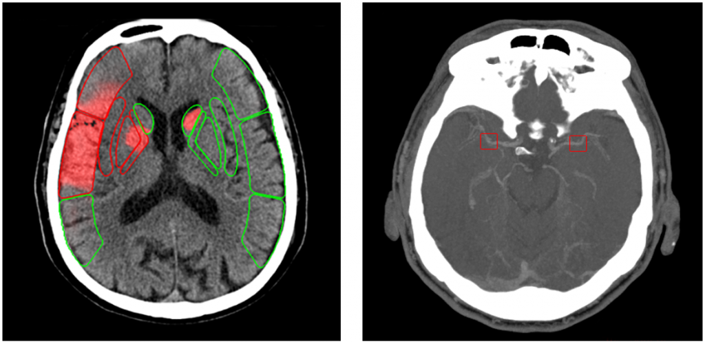
AUTOStroke from Canon Medical integrates a comprehensive set of stroke applications including: Non-contrast CT Intracranial Haemorrhage that helps quickly locate multiple variations of intracranial haemorrhage when time is critical; Non-contrast CT ASPECTS delivering scoring of early ischemic stroke signs that are routinely challenging to detect; CT Large Vessel Occlusion to help locate occlusions in cerebral infarction patients; and CT Perfusion maps created using validated Bayesian CTP+ algorithms to help indicate potential regions of penumbra and infarct core.

Photo caption: AUTOStroke integrates a comprehensive set of stroke applications including (1) Non-contrast CT ASPECTS delivering scoring of early ischemic stroke signs that are routinely challenging to detect; and (2) CT Large Vessel Occlusion to help locate occlusions in cerebral infarction patients.


Three-minute Chest X-ray test using AI producing encouraging results

An Artificial Intelligence (AI) programme created by Bering Limited and study conducted by iCAIRD, Scotland’s Industrial Centre for AI Research in Digital Diagnostics has yielded promising results from Chest X-rays in a simulated clinical test setting to speed up Covid-19 diagnosis in patients who had presented to hospital Emergency Departments (ED) with respiratory symptoms.

The iCAIRD studyi, funded by Innovate UK, was in partnership with NHS Greater Glasgow and Clyde, using Canon Medical Research Europe’s Safe Haven Artificial Intelligence Platform (SHAIP) as well as datasets from the Glasgow Safe Haven. It used a new AI algorithm giving an accurate Covid-19 result in a test environment under three minutes with performance on par with four certified radiologists.

“This is another welcome development demonstrating the potential for AI to support clinicians ensuring patients are getting the highest quality and most relevant treatment,” states Prof David Lowe, Joint Clinical Lead of the West of Scotland Innovation Hub and an Emergency Medicine Consultant at Queen Elizabeth University Hospital. “Through testing in a safe environment we have been able to see that this algorithm can identify Covid-19 on Chest X-rays that were routinely taken during initial clinical assessment. This could not just help with the treatment of patients but may also speed up the process of isolating infected patients.”

Dr Mark Hall, Radiology Consultant at NHS Greater Glasgow & Clyde added, “We continue to see the positive potential impact AI could have on radiology, from reducing waiting times to improving accuracy and reducing pressures on staff. Ongoing research highlights the importance of using developments in AI to enhance diagnosis and treatment. The level of accuracy may allow consultants to make even more informed decisions as we have a greater pool of data to use. There can often be a misconception that AI input will mean the public gets less time with doctors, but this is not the case. Technology like this may help us speed up processing high numbers of similar cases, while retaining accuracy, allowing for more time with patients and more complex cases.”

“Covid-19 along with many chronic diseases continually put pressure on our UK health services. Research and development into how we can speed-up diagnostic imaging is therefore incredibly important,” states Mark Hitchman, Managing Director of Canon Medical Systems UK. “There are also broader benefits of having AI research situated in the UK via our sister company Canon Medical Research Europe. It means that the AI algorithms developed using the Safe Haven Artificial Intelligence Platform are specific in terms of demographics, meaning more readiness for UK patient population deployment.”

The Canon Medical Research Europe AI Centre of Excellence includes a team of data sciences, clinical analysts and software engineers based in Edinburgh who collaborate with the universities of Glasgow and Aberdeen and NHS hospitals including Queen Elizabeth University Hospital Glasgow and Aberdeen Royal Infirmary. The team is developing a set of tools to help clinicians to create novel AI solutions using UK patient data for machine learning, together with infrastructure for data scientists to develop, train and validate algorithms without patient data ever leaving the hospital environment.

“Collaboration with academia, NHS and industry is vital for the safe collection of usable data, its annotation and testing, plus deployment into active AI research,” states Ken Sutherland, President of Canon Medical Research Europe. “Our work in Edinburgh underpins many exciting AI research projects in automation, decision support and ultimately precision medicine. We recently celebrated our 100th patent and are proud to play a key role in advancing NHS access to the exciting opportunities of AI.”

i. Drozdov, I., Szubert, B., Reda, E. et al.Development and prospective validation of COVID-19 chest X-ray screening model for patients attending emergency departments. Sci Rep11, 20384 (2021). https://doi.org/10.1038/s41598-021-99986-3

Photo Caption: Image courtesy of iCAIRD’