Deep Learning Spectral
A fully integrated end-to-end spectral workflow
The Aquilion ONE / PRISM Edition harnesses the temporal benefits of rapid kV switching with patient specific mA modulation and combines them with a Deep Learning Reconstruction that delivers excellent energy separation and low-noise properties.
What’s more, its fully integrated end-to-end workflow is easy to use and can be conveniently incorporated into your routine protocols.
Whitepaper
Deep Learning Spectral CT
– Faster, easier and more intelligent
Kirsten Boedeker, PhD, DABR, Senior Manager, Medical Physics*1
Mariette Hayes, Global CT Education Specialist, Healthcare IT*1
Jian Zhou, Senior Principal Scientist*2
Ruoqiao Zhang, Scientist*2
Zhou Yu, Manager, CT Physics and Reconstruction*2
*1 Canon Medical Systems Corporation
*2 Canon Medical Research USA
a full 160 mm of coverage
rapid kv switching with patient specific ma modulation
spectral deep learning reconstruction
a seamless workflow
New spectral applications integrated with the Aquilion ONE / PRISM Edition
Images can be delivered directly to your reading station, and with our range of new Vitrea applications, you can easily analyze comprehensive spectral data, including quantification and multi-layered images for a more detailed and definitive diagnosis.
Smartly developed, these applications provide a seamless solution from scan to diagnosis, and are scalable from a single workstation to a hospital-wide network, including PACS integration.
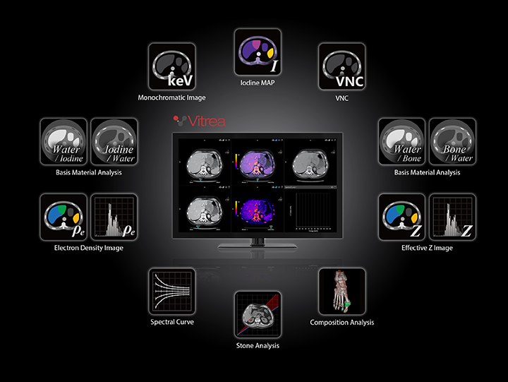
Vitrea Advanced Visualization *
Expand your spectrum with our latest healthcare IT solutions
Review Spectral data quickly and easily with a range of applications that simplify your workflow and provide clinically relevant features and outputs for every patient.
- Create effective monochromatic images for 166 energy levels ranging from 35 keV to 200 keV.
- View quantitative color-coded iodine distribution maps that show the iodine uptake in anatomical structures.
- Characterize and differentiate tissue with effective Z and Electron density information at your fingertips.
- Differentiate between uric acid and non-uric acid urinary stones to make better-informed treatment decisions.
- Visualize and quantify the presence of monosodium urate in anatomical structures.
- Differentiate iodine from an intracranial hemorrhage.
View Spectral Clinical Gallery
* Vitrea is available as an option.
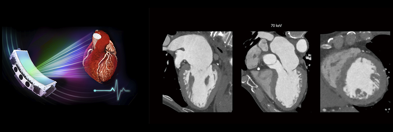
ECG gated volumetric Deep Learning Spectral cardiac CT
Technological Advantages
- 16 cm volumetric coverage
- One beat cardiac acquisition
- SEMAR
- Temporal congruency
- Spatial alignment
Clinical advantages
- Low keV to improve iodine conspicuity and reduce iodine contrast load
- Myocardial perfusion analysis
- Delayed enhancement analysis