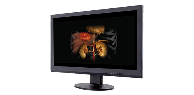
Imaging | Computed Tomography
Our CT innovations are designed for accurate oncology imaging with simplified workflows, high spatial resolution and automated dose reduction.
CT City Hopper
Clinical Confidence Anywhere
With screening programs becoming more important in today’s challenge to find cancer in an early stage, the question is, how to get as many as possible eligible people scanned in an easy way.
The CT City Hopper is Canon Medical’s Mobile Imaging Solution that is most flexible moving from location to location, an ideal addition to support a cancer screening project.


Dr. Santiago Viteri, MD
Medical Oncologist and Patient Partner
UOMi Cancer Center, Barcelona, Spain
Click here to watch the full webinar
Prof. Hannah Hazard-Jenkins, MD, FACS
Associate Professor of Surgery at the department of Surgery WVU School of Medicine
Director of the WVU Cancer Institute
Morgantown, West Virginia, USA
Click here to watch the full webinar

Aquilion Precision
Precision in Every Detail
Introducing the world’s first Ultra-High Resolution CT (UHR CT), capable of resolving anatomy as small as 15 microns while delivering the image resolution of a catheterization laboratory.
Overview Video

Ultra-High Resolution CT
- UHR Detector
- UHR DAS
- UHR Tube
- UHR Gantry
- UHR Reconstruction
0.25 mm
Detector
150 micron
Spatial Resolution
50 lp/cm at 0% MTF
Reference only

1024 x 1024 Matrix
Aquilion Precision UHR CT
Whitepaper
Kirsten Boedeker, PhD, DABR Senior Manager, Quantitative Image Quality, Canon Medical Systems Corporation
New UHR Reconstruction Matrix
Detector


UHR Detector


Clinical Example
Lung1
SHR / 0.25 mm
Courtesy of Kyorin University, Japan


Lung2
HR / 0.5 mm
Courtesy of Prof. Prokop RadboudUMC, Nijmegen, the Netherlands


Improving Patient Outcomes
Can UHR CT Improve Outcomes with Better Detection, Diagnosis and Staging of Pancreatic and Biliary Cancer?
Miyuki Sone, M.D.
National Cancer Center Hospital,
Tokyo, Japan

Aquilion Prime SP
Oncology Care with a Streamlined Workflow
Built with premium technology migrated straight from our high-end CT, Aquilion Prime SP empowers you to care for all patients, from pediatric to bariatric, while providing your team with the fast, efficient solutions they need.
System Highlights:
- Up to 4 cm z-axis coverage
- 0.5 mm × 80 PUREViSION detector row
- 0.35 sec rotation

PUREViSION Optics
Aquilion Prime SP strikes the perfect balance between image quality and dose – transforming routine imaging with new levels of detail, resolution and safety.


Whitepaper
Jay Vaishnav, PhD Clinical Scientist, Manager, Medical Affairs, Canon Medical Systems USA, Inc.
vHP3*—Variable Helical Parameters
Overall faster scan times make vHP3 compliant to the needs of the patient, and shorten exam times for trauma imaging when every second is critical.
With this technology, a single series reconstruction enables several studies to be interpreted simultaneously for faster scans when every second counts.
vHP3 may also help you:
- Decrease radiation dose
- Decrease the amount of IV contrast needed
- Improve the workflow of complex clinical exams
*Option

Easy Patient Positioning
Aquilion Prime SP’s accessible gantry and couch have been designed for enhanced comfort, particularly for bariatric patients or those who are elderly and living with cancer.
*Option

Aquilion Prime SP’s accessible gantry design and powerful advanced imaging applications deliver an ideal oncology solution to assist clinicians with visualization, staging and treatment planning.

Adaptive Iterative Dose Reduction 3D

Single Energy Metal Artifact Reduction (SEMAR)

SURESubtraction unique Differential Iodine Enhancement
Aquilion ONE / PRISM Edition
Transforming CT and Oncology care
Clinically-focused research and technological developments have culminated in a CT system that reduces dose and simplifies workflows for tumor visualization and assessment.
System Highlights:
- Up to 16 cm z-axis coverage
- 0.5 mm × 320 PUREViSION detector row
- 0.275 sec rotation

SilverBeam Filter
The detail of a CT at the dose on the order of an X-ray exam
SilverBeam, a beam shaping energy filter, leverages the photon-attenuating properties of silver to selectively remove low energy photons from a polychromatic X-ray beam, leaving an energy spectrum optimized for lung cancer screening.
Deep Learning Spectral
A fully integrated end-to-end spectral workflow
The Aquilion ONE / PRISM Edition harnesses the temporal benefits of rapid kV switching with patient specific mA modulation and combines them with a Deep Learning Reconstruction that delivers excellent energy separation and low-noise properties.
What’s more, its fully integrated end-to-end workflow is easy to use and can be conveniently incorporated into your routine protocols.

Tumor Assessment with Whole Organ Perfusion
Volume Perfusion Liver*
With the ability to evaluate tumor response or anti-angiogenesis, CT body perfusion has become a valuable clinical tool – especially now that you’ve got 16 cm of z-axis coverage at your disposal.
Liver Perfusion Protocol: DLP = 720.8 mGy.cm, Effective dose = 10.81 mSv (k = 0.015)
*CT Volume perfusion only available with Aquilion ONE

Outstanding Lesion Visualization and Assessment
SURESubtraction Body
- Assessment of tumors, lymph nodes and vascular lesions
- Infarcts, ischemia
- Blood flow mapping
- Visualize local perfusion differences
- Display local iodine concentration
- Visualize contrast enhancement

Expert Tools
Learn More CT Clinical Applications >
* CT Volume perfusion only available with Aquilion ONE
Imaging | Magnetic Resonance
Our MRI solutions provide optimized workflow and image quality to help you deliver efficient and effective care to each of your oncology patients.
Vantage Galan 3T
Optimized Image Quality and Patient Comfort
Get streamlined workflow, high-quality images and maximum patient comfort with Canon Medical’s Vantage Galan 3T MR system. This system has been designed to meet the needs of your team and your patients with maximum comfort, image quality and workflow efficiency.
Deep Learning Reconstruction, AiCE
- Applicable to 96% of MRI procedures*, Advanced intelligent Clear-IQ Engine (AiCE) removes noise and increases SNR to help you to see through the noise to deliver clear, sharp and distinct images
- Combined with accelerated scan technologies, AiCE intelligently expands your capabilities to help reduce time while still producing stunning images
- Advanced post processing capability with Olea/Vitrea technologies enhances intelligent diagnostic decision making
Unique Acoustic Noise Reduction Suites
Pianissimo Zen¹
*99% reduction by unit of Loudness level “dB” and 97% reduction by unit of perceived loudness “Sone”
¹ Optional

Whole body DWI
We’ve evolved our MRI solutions to deliver wide-range, low-distortion DWI so you can identify tumors and lesions anywhere in your patient’s body.
Whole body image with WFS DIXON
WFS* DIXON achieves consistent fat suppression and homogeneity while acquiring four different tissue contrasts in one scan, reducing the total number of scans needed.
Available for T1, T2, and PD sequences, WFS DIXON can be acquired in any area of the body.
* WFS: Water Fat Separation

Quick Star
Quick Star free-breathing and motion reduction is an incredibly helpful tool for examining patients that have difficulty holding their breath – especially when conducting liver exams or scanning pediatric patients.

Post-processing & 3D Visualization
Tumor Assessment
Our image processing software solutions help you view and analyze a range of tumor characteristics, including volume, cellularity (DWI), aggressiveness (DCE), and perfusion (DSC/IVIM).

*Designed and manufactured by Olea Medical
Applications
– Lesion analysis*
– Longitudinal analysis*
– Brain perfusion*
– IVIM*
Applications
– Multi-parametric analysis*
Applications
– Breast MR
– BI-RADS reporting tools*
– Breast tumor follow-Up*
Applications
– Computed DWI (cDWI) maps
– Multi-parametric analysis
– PI-RADS reporting tools*
– IVIM*
Whitepapers
Borderline Ovary Tumor
(Borderline serous cystadenoma)
Prostate Adenocarcinoma
Lesion Analysis
Longitudinal Analysis
IVIM—Intravoxel Incoherent Motion
Quantitative separation of diffusion and perfusion from multi b-value DWI
Prostate Cancer—Multi-Parametric Analysis
PI-RADS Reporting Tools
– Characterization
– Localization
– Grading/Staging
Breast Cancer
BI-RADS Reporting Tools
Imaging | Ultrasound
Canon Medical’s comprehensive Aplio i-series provides unprecedented resolution and penetration for quick and reliable diagnoses.
Aplio i-series / Prism Edition
Intuitive. Intelligent. Innovative.
Crystal-clear images and expert tools deliver outstanding clinical precision and departmental productivity.
Innovative System Architecture:
- iBeam Forming
- intelligent Dynamic Micro-Slice (iDMS)
- Multiplexing Architecture
- Transducer Technology

Comprehensive Experts Tools
*Availability dependent on system

Superb Micro-vascular Imaging (SMI)* offers enhanced flow visualization.

MicroPure provides a better view of microcalcification in breast tissue.

Precision Imaging offers better border definition and clarity of structures.

Contrast Enhanced Ultrasound Suite (CEUS) lets you assess liver lesions and utilize CEUS LI-RADS® categorization.

Shear Wave Elastography* for quantitative assessment of tissue stiffness and Shear Wave Dispersion* for assessment of dispersion slope which is a property related to tissue viscosity leading to more confident diagnosis.

Quad View* lets you see live ultrasound images from multiple modes, as well as those fused with volume data to help you identify lesions more confidently.
Accurate Pre And Post-therapy Cardiac Function Assessment
2D and 3D* Cardiac Wall Motion Tracking
Aplio’s advanced Wall Motion Tracking technology provides immediate visual and quantitative access to global and regional myocardial wall motion dynamics (in 2D and 3D) on both live and previously stored images.
*Availability dependent on system


